Clown Fish
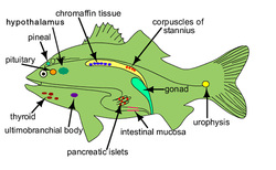
Adrenal Gland
There is no true adrenal gland present in most fish (exception is sculpins). The adrenal cortical tissue in most fish is represented by the interrenal cells. These cells are pale eosinophilic cuboidal cells associated with major blood vessels in the anterior kidney. Both glucocorticoid and mineralocorticoid are secreted.
The adrenal medullary cells (chromaffin cells) may vary in location. These cells are usually found with the sympathetic ganglia in clumps between the anterior kidney and spine or in the interrenal tissue.
Thyroid Gland
The thyroid follicles are very similar to mammalian thyroid tissue. Thyroid follicles are distributed throughout the connective tissue of the pharyngeal area and may be observed around the eye, ventral aorta, hepatic veins and anterior kidney. It is important to realize that thyroid tissue can be widely distributed. Many times pathologist have erroneously considered this distribution of normal thyroid tissue to represent metastasis from a thyroid follicular cell tumor.
Endocrine Pancreas
The endocrine pancreas is present in most fish as islet of Langerhans and is associated with the exocrine pancreas. In some species the islets are very large and may be grossly visible (Brockman bodies). During the spawning season the size and number of islet will increase in some fish. These should not be confused with an adenoma.
Parathyroid Glands
The parathyroid glands are absent in fish; their function is taken over by other endocrine organs. (Corpuscles of Stannius)
Ultimobranchial Gland
This gland lies ventral to the esophagus in the transverse septum separating the heart from the abdominal cavity. This organ secretes calcitonin (lowers serum calcium levels) that acts with hypocalcin (secreted by the corpuscles of Stannius) to regulate calcium metabolism.
Corpuscles of Stannius
These are islands of eosinophilic granular cells located in paired organs on the ventral surface of the kidney. This organ secretes a protein called hypocalcin (teleocalcin) that acts with calcitonin to regulate calcium metabolism.
Urophysis
This is a neurosecretory organ found on the ventral aspect of the distal end of the spinal cord. These bodies are composed of unmyelinated axons terminating on a capillary wall. The function of the urophysis is unknown.
Pineal Gland
The pineal gland is a light sensitive neuroendocrine structure that lies in the anterior brain and is a well-vascularized organ. This gland secretes melatonin that may play a role in controlling reproduction, growth, and migration.
